Signature Healthcare
Conventional ultrasound displays the images in thin, flat sections of the body. Advancements in ultrasound technology include three-dimensional (3-D) ultrasound that formats the sound wave data into 3-D images. Ultrasound examinations do not use ionizing radiation (as used in x-rays), thus there is no radiation exposure to the patient. Because ultrasound images are captured in real-time, they can show the structure and movement of the body's internal organs, as well as blood flowing through blood vessels. Doctors use ultrasound to detect changes in the appearance of organs, tissues, and vessels and to detect abnormal masses, such as tumors.
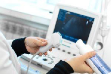
Ultrasound is safe and painless. Ultrasound imaging, also called ultrasound scanning or sonography, involves the use of a small transducer (probe) and ultrasound gel placed directly on the skin. The transducer is a small hand-held device that resembles a microphone. High-frequency sound waves are transmitted from the probe through the gel into the body. Some exams may use different transducers (with different capabilities) during a single exam. The transducer sends out inaudible, high-frequency sound waves into the body and listens for the returning echoes. When a sound wave strikes an object, it bounces back or echoes. By measuring these echo waves, it is possible to determine how far away the object is as well as its size, shape, and consistency. This includes whether the object is solid or filled with fluid. Ultrasound imaging uses the same principles as the sonar that bats, ships, and fishermen use.
At Signature Healthcare, we offer Sonohysterography, also known as saline infusion sonography, which is a special, minimally invasive ultrasound technique. It provides pictures of the inside of a woman’s uterus.
How should I prepare?
Wear comfortable, loose-fitting clothing. You may need to change into a gown and remove jewelry in the area to be examined.
Preparation for the procedure will depend on the type of exam you will have. For some scans, your doctor may tell you not to eat or drink for up to 12 hours before your exam. This timeframe is lower for babies and young children. For others, the doctor may ask you to drink up to six glasses of water two hours prior to your exam and avoid urinating. This will ensure your bladder is full when the scan begins.
What should I expect during the procedure?
The technologist applies a small amount of gel to the area under examination and places the transducer there. The gel allows sound waves to travel back and forth between the transducer and the area under examination. The ultrasound image is immediately visible on a video monitor. The computer creates the image based on the loudness (amplitude), pitch (frequency), and time it takes for the ultrasound signal to return to the transducer. It also considers what type of body structure and/or tissue the sound is traveling through.
How long does the procedure take?
Most ultrasound exams take about 30 minutes. More extensive exams may take up to an hour.
Children/Family members
For safety reasons, children are not allowed in the exam rooms. If you must bring your child, please bring someone to watch them in the waiting room. For OB exams, family members may have access to the exam room AFTER the diagnostic portion of the exam is completed.
Getting Your Results
Contact the ordering physician or visit your MyHealth Portal for your results.