Signature Healthcare
When very small blood vessels just below the skin’s surface become damaged, they can form webs of blue, purple or red veins. Spider veins, a milder type of varicose veins, are smaller than varicose veins and often look like a sunburst or "spider web." These "spider veins" rarely cause serious symptoms, but because they are near the surface of the skin, their blue or red color is visible.
Varicose veins are enlarged veins visible through the skin and may appear as blue or purple twisted, knot-like cords. Varicose veins can occur anywhere in the body, but are more commonly found on the legs. Hemorrhoids, a type of varicose vein, can appear during pregnancy around the anus or in the vagina.
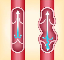
Varicose veins are caused by increased blood pressure inside the superficial leg veins. Superficial veins are near the surface of the skin, whereas deep veins are located in the muscle tissue. In contrast, deep veins lead to the vena cava, a large vein that transports blood to the heart.
The blood in the veins of the legs works against gravity in order to return upwards to the heart by one-way valves in the veins. When the leg muscles contract and squeeze the deep veins, the valves inside the veins open. When the leg muscles relax, the valves close, preventing blood from flowing backward.
When the one-way valves become weakened or damaged, blood can collect in the veins, causing the veins to become enlarged. Sitting or standing for long periods can cause blood to pool in the leg veins, increasing the pressure within the veins. In persons who are prone to varicose veins, the veins can stretch as a result of increased pressure. This stretching of the veins may weaken the walls of the veins and damage the valves. Thick varicose veins or spider veins may result.
While varicose veins are not considered a severe medical condition, they can be uncomfortable and can lead to more serious problems such as phlebitis (inflammation in the leg) or blood clot. Varicose veins can also be a cosmetic concern to some people.
With severe varicose veins, there is a small increased chance of developing deep vein thrombosis(DVT). DVT requires immediate medical attention. Symptoms of DVT include sudden, severe leg swelling and can result in blood clots that travel to the brain or the heart.
Each individual may experience symptoms differently.
Severe varicose veins may eventually produce long-term leg swelling resulting in more serious skin and tissue problems, such as ulcers and non-healing sores.
About 15% of adults in the U.S. have varicose veins. The risk of varicose veins is strongly related to age and gender.
Obesity
Obesity is a major risk factor for varicose veins. Excessive weight increases the pressure on the veins of the legs and aggravates the condition.
Family History
Heredity is important in determining susceptibility to varicose veins, but the specific factors responsible for this have not been identified.
Inactivity
Prolonged standing or sitting increases pressure in the veins.
Gender
Women are particularly susceptible to varicose veins because of the influence of progesterone on the veins and the effects of pregnancy. Women are 2-3 times more likely to have varicose veins.
Pregnancy
Pregnant women have an increased risk of developing varicose veins due to the hormonal influences of pregnancy on the veins, but the veins often return to normal within one year of childbirth. Women who have multiple pregnancies may develop permanent varicose veins.
Age
Varicose veins usually affect people between the ages of 30 and 70. With advancing age, the elastic shell of the vein begins to weaken increasing the chance that the vein will dilate.
In addition to a complete medical history and physical examination, diagnostic procedures for varicose veins may include any or a combination of the following:
Duplex Ultrasound
A type of vascular ultrasound procedure done to assess blood flow and the structure of the leg veins. The term "duplex" refers to the fact that two modes of ultrasound are used - Doppler and B-mode. The B-mode transducer (like a microphone) obtains an image of the vessel being studied. The Doppler probe within the transducer evaluates the velocity and direction of blood flow in the vessel.
Color-Flow Imaging
(Also called triplex ultrasound.) A procedure similar to duplex ultrasound that uses color to highlight the direction of blood flow. Vessels in which blood is flowing are colored red for flow in one direction and blue for flow in the other, with a color scale that reflects the speed of the flow.
Magnetic Resonance Venography (MRV)
A diagnostic procedure that uses a combination of a large magnet, radiofrequencies, and a computer to produce detailed images of organs and structures within the body. An MRV uses magnetic resonance technology and intravenous (IV) contrast dye to visualize the veins. Contrast dye causes the blood vessels to appear opaque on the x-ray image, allowing the physician to visualize the blood vessels being evaluated. MRV is useful in some cases because it can help detect causes of leg pain other than vein problems.