Signature Healthcare
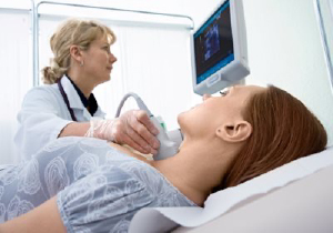
The Signature Healthcare Non-Invasive Vascular Laboratories specialize in the detection and diagnosis of vascular (arteries/veins) related diseases by using state-of-the-art ultrasound and physiological testing equipment performed by a Registered Vascular Technologist.
Both sites are fully accredited by the Inter-Societal Commission for the Accreditation of Vascular Laboratories (ICAVL), the premier international accreditation body in the industry. Signature Healthcare Vascular Laboratory Services are committed to providing the highest of patient care and quality of testing.
Signature Healthcare Vascular Laboratory Services proudly offers the following exams on an outpatient basis.
Carotid and Vertebral Artery Scan
The carotid arteries are located on each side of the neck and they supply your brain with oxygenated blood. The vertebral arteries are located in the back of your neck, and they too, supply oxygenated blood flow to your brain. Over time the carotid arteries can narrow, due to plaque formation, which may reduce the blood flow to the brain with potential break-up of plaque, resulting in small fragments of plaque to travel towards the brain, which can cause stroke and TIAs (Transient Ischemic Attacks). Your arteries are normally smooth and unobstructed on the inside. However, as you age, a sticky substance called plaque can build up in the walls of your arteries. Plaque is made of cholesterol, calcium and fibrous tissue. As more plaque builds up, your arteries narrow and stiffen. This process is called atherosclerosis, or hardening of the arteries. Carotid artery disease is a serious health condition, because it is the leading source for stroke. Some of the risk factors that can be associated with the progression of atherosclerosis are smoking, high blood pressure, high cholesterol and diabetes. Unfortunately, the first sign of carotid artery disease could be a stroke. However, you may experience warning symptoms of a stoke called transient ischemic attacks or TIAs. These symptoms usually last for a few minutes to an hour. These symptoms may include a feeling of weakness or numbness (usually affected on one side of the body), slurred or gargled speech, or a vision loss or defect, usually affecting only one eye.
Venous Duplex Ultrasound Scanning
Vascular diseases affecting the veins is more common with symptoms such as leg pain, swelling, warmth and redness/inflammation of the skin and visible varicose and spider veins. Venous Duplex ultrasound can be performed to detect superficial vein thrombosis and damaged or leaking valves that may be the result of varicose veins within the venous system. Varicose veins are caused by increased blood pressure inside the superficial (close to the skin surface) vein(s) due to the presence of weakened or damaged one-way valves in the veins. Deep Vein Thrombosis (DVT) is the formation of a blood clot in the deep vein of the leg. DVT can cause symptoms of leg swelling and pain. In the event that a clot breaks away and travels through the blood stream to block blood vessels of the lungs (pulmonary embolism), it can lead to severe difficulty breathing and even death, depending on the degree of blockage. In the United States, about 2 million people annually develop DVT. Most of them are aged 40 years or older. Statistics reveal that at least 200,000 patients die each year from blood clots in their lungs.
AortoIliac Duplex Scan
The abdominal aorta is the largest artery in the abdomen that branches into the iliac arteries in the lower abdomen to the right and left sides of the pelvis. The aorta also has branches that supply blood flow to major organs in the body. Abdominal aortic aneurysms (bulge or enlargement of a weakened area in the arterial wall) affect over 2.3 million Americans, but rarely produce symptoms, unless they rupture. If the aneurysm should rupture, there is a mortality rate making aneurysms the 13th largest cause of death in the United States. In addition, blockages in the abdominal aorta and iliac (located in pelvis) arteries may leave a patient with pain in the hip, buttocks, or thigh muscles during exercise. An ultrasound study can detect these abnormalities.
Renal Artery Duplex Ultrasound Scanning
A build-up of plaque in the renal arteries (blood vessels supplying flow to the kidneys) can cause narrowing that restricts blood flow to the kidneys. This can lead to kidney failure or could be the reason for a patient's hypertension (high blood pressure). Renal Artery Ultrasound can be performed to detect narrowing in the renal arteries, blood flow in the kidneys and evaluate the kidney organ itself for any abnormalities.
Lower and Upper Extremity Arterial Testing
Approximately 12 million Americans suffer from Peripheral Arterial Disease (PAD) - hardening of the arteries, or atherosclerosis. This narrowing of the blood vessels in the legs is caused by a build-up of plaque in the arteries. Patients with PAD have a much higher incidence for heart attack and stroke. Testing with blood pressure measurements of the legs and hand held Doppler ultrasound probes can be used to identify the presence of narrowing or blockages in the arteries, its location and severity.
Arterial Indirect Physiological Arterial Testing of the Upper/Lower Extremities
Evaluates for PAD. Peripheral Vascular Disease with PVR’s, Doppler Waveform Analysis and Segmental Blood Pressure Measurements.
Arterial Duplex Scanning
Upper/Lower Extremities, including native arteries and arteries that have been surgically repaired or reopened.
Bypass Graft Duplex Scanning
Upper/Lower Extremities for the evaluation of patients who have had a bypass graft placed in their leg or arm.
Hemodialysis Access Duplex Scanning
Evaluates Arteriovenous fistulas or Arteriovenous grafts that have been placed in the patients arm or leg. This is seen in patients whose kidneys have failed.
Mesenteric Duplex Scanning
Evaluates the blood vessels associated with the digestive tract.
Hepatoportal Duplex Scanning
Evaluates the circulation of blood flow to the liver from digestive organs, spleen and pancreas.
Temporal Artery Duplex Scanning
Located at each side of head, evaluates the patient with suspected Temporal Arteritis, prior to a Temporal Artery Biopsy.
